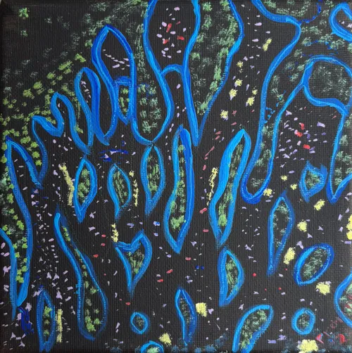
Intestine
15x15 · acrylic on canvas · 2025
Fluorescence microscopy image of an intestinal tissue sample from a Crohn’s disease patient. Red shows T cells, green marks a specific subset of T cells of interest, blue indicates cell nuclei, and yellow represents the overlap of red and green signals.
Protuhlo